- HOME
- Vienna 2024
- Lithuania 2023
- 16th CEEPC Prague 2022
- Proteome & Proteomics
- Proteomics Potentiality
- Precision Medicine & Cancer
- Proteomics and COVID-19
- Big Data & AI
- Spotlight Lithuania
- Humanity
- Meeting Reports & Tributes
- Country Profiles
- Proteomic Snippets
- Enabling Advances
- Spotlight Czech Republic
- Spotlight Poland
- 13th CEEPC - Ustron, Poland
- Spotlight Romania
- 12th CEEPC - Bucharest
- Spotlight Slovakia
- Spotlight Macedonia
- Sports Medicine
- Contacts & Copyrights
Proteomic Snippets
Proteomics is a systems approach for the global study of protein expression changes. It can provide information on gene function, disease processes and mechanisms of drug action at several stages in the drug discovery pipeline and pave the way for improved and faster implementation of drug discovery strategies.
Since proteomics encompasses a number of multi disciplines, it has a major role to play in our understanding of biological processes and diverse diseases.
 Lecanemab in Early Alzheimer’s Disease - November 29, 2022
DOI: 10.1056/NEJMoa2212948
Christopher H. van Dyck, M.D., Chad J. Swanson, Ph.D., Paul Aisen, et al. - New England Journal of Medicine
The accumulation of soluble and insoluble aggregated amyloid-beta (Aβ) may initiate or potentiate pathologic processes in Alzheimer’s disease. Lecanemab, a humanized IgG1 monoclonal antibody that binds with high affinity to Aβ soluble protofibrils, is being tested in persons with early Alzheimer’s disease. Lecanemab reduced markers of amyloid in early Alzheimer’s disease and resulted in moderately less decline on measures of cognition and function than placebo at 18 months but was associated with adverse events. Longer trials are warranted to determine the efficacy and safety of lecanemab in early Alzheimer’s disease.
The next horizon in precision oncology: Proteogenomics to inform cancer diagnosis and treatment. DOI:https://doi.org/10.1016/j.cell.2021.02.055. Henry Rodriguez, Jean Claude Zenklusen, Louis M. Staudt , James H. Doroshow & Douglas R Lowy. Cell, Vol. 184, April 1, 2021
Proteogenomics may provide better prospects to the clinical characterization of tumors, help make a more accurate diagnosis of cancer, and improve treatment for patients with cancer. In precision oncology, proteogenomics needs to be fully integrated into clinical trials and patient care in order for precision oncology to deliver the right cancer treatment to the right patient at the right dose and at the right time.
Ageing issues and Arthritis - does protective signalling malfunctions during ageing?
Credit: L. Bryan Ray Science 2020;370:1429
Science 18 Dec 2020: Vol. 370, Issue 6523, pp. 1429, DOI: 10.1126/science.370.6523.1429-a
A signaling connection that allows transforming growth factor–β (TGF-β) to protect cartilage from osteoarthritis could guide new therapeutic strategies. Expression of the transcription factor FoxO1 in cartilage decreases with aging. Loss of FoxO family members causes osteoarthritis-like damage in mice and humans. Wang et al. found that TGF-β specifically promotes expression of the Fox01 isoform in isolated mouse chondrocytes in a manner that depends on activation of TGF-β–activated kinase 1 (TAK1). Overexpression of FoxO1 in mice protected against experimentally induced arthritis. The protective TGF-β/TAK1 signaling nexus appeared to act, at least in part, by activating autophagy.
Search-and-replace genome editing without double-strand breaks or donor DNA.
Nature (2019) Published: 21st October 2019 - https://doi.org/10.1038/s41586-019-1711-4,
Andrew V. Anzalone, Peyton B. Randolph, Jessie R. Davis, Alexander A. Sousa, Luke W. Koblan, David R. Liu et al .........
The gene-editing tool CRISPR is, in reality, pretty hard on the genome. It’s a pair of DNA scissors that cuts the double helix, and what’s called “editing” is actually a cell’s hasty attempt to patch things back together. That introduces errors. Dr David Liu, a Harvard University biologist, together with his team has come up with recent improvements to CRISPR technology by introducing “prime editing,” a molecular gadget he says can rewrite any type of genetic error without actually severing the DNA strand, as CRISPR does.
The new technology uses an engineered protein that can transform any single DNA letter into any other, as well as add or delete longer stretches. Researchers claim it’s capable of repairing nearly any of the 75,000 known mutations that cause inherited disease in humans.
CRISPR 1.0 is harnessed most often to disable genes—making it useful for research and possibly in treating a subset of diseases where a DNA delete button is what’s called for. More extensive gene replacements are also possible with this tool but aren’t easy to control.
The promise to potentially resolve the entire spectrum of inherited human ailments is huge, but in practice it’s still distant. The prime editor is, in molecular terms, gigantic— so getting it into people’s cells is going to require something like gene therapy.
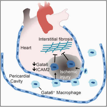
Gata6+ Pericardial Cavity Macrophages Relocate to the Injured Heart and Prevent Cardiac Fibrosis.
Justin F. Deniset, Darrell Belke, Woo-Yong Lee, Selina K. Jorch, et al. Immunity, 16th July, 2019; 51 (1): 131 DOI: 10.1016/j.immuni.2019.06.010
Macrophages play an important role in structural cardiac remodeling and the transition to heart failure following myocardial infarction (MI). This study examined the contribution of resident cavity macrophages located in the pericardial space adjacent to the site of injury. They found that disruption of the pericardial cavity accelerated maladaptive post-MI cardiac remodeling. Gata6 + macrophages in mouse pericardial fluid contributed to the reparative immune response.
Gata6 + macrophages were present in human pericardial fluid, supporting the notion that this reparative function is relevant in human disease and uncovers an immune cardioprotective role for the pericardial tissue compartment and argue for the reevaluation of surgical procedures that remove the pericardium.
Discovery of a new cell that can help heal injured heart muscle may open the door to new therapies and hope for the millions of people who suffer from heart disease. We always knew that the heart sits inside a sac filled with a strange fluid. Now we know that this pericardial fluid is rich with healing cells. These cells may hold the secret to repair and regeneration of new heart muscle.
Exosomes from mesenchymal stem/stromal cells: a new therapeutic paradigm
Kan Yin et al. Biomarker Research20197: https://doi.org/10.1186/s40364-019-0159-x , Published: 4 April 2019
Mesenchymal stem/stromal cells (MSCs) have been demonstrated to hold great potential for the treatment of several diseases. Their therapeutic effects are largely mediated by paracrine factors including exosomes, which are nanometer-sized membrane-bound vesicles with functions as mediators of cell-cell communication. MSC-derived exosomes contain cytokines and growth factors, signaling lipids, mRNAs, and regulatory miRNAs. Increasing evidence suggests that MSC-derived exosomes might represent a novel cell-free therapy with compelling advantages over parent MSCs such as no risk of tumor formation and lower immunogenicity. This paper reviews the characteristics of MSC exosomes and their fate and highlights the therapeutic potential of MSC-derived exosomes in liver, kidney, cardiovascular and neurological disease.
Co-regulatory networks of human serum proteins link genetics to disease. Valur Emilsson et al.
Science 24 Aug 2018: Vol. 361, Issue 6404, pp. 769-773
DOI: 10.1126/science.aaq1327
Detecting such interacting networks of proteins within the complex proteome of the blood requires enormous amounts of detailed data. Access to medical information, biometric data, and blood samples of 5,457 Icelanders who were part of a Reykjavik-based aging study called Age, Gene/Environment Susceptibility (AGES). This team was able to design a panel of DNA aptamers—short unique sequences that bind specific proteins—capable of recognizing and measuring 4,137 known and predicted serum proteins within the subject samples. Together, this enabled “the largest proteomic analysis of secreted proteins to date,”
“Identification of the human skeletal stem cell,” C.K.F. Chan et al., Cell, doi:10.1016/ j.cell.2018.07.029, 2018.
The researchers started with a human fetal femur and sorted the nonhematopoietic cells from blood precursors. They then sequenced individual, nonblood cells’ RNA. Based on the expression profiles of cells located in areas of active growth in the fetal bone, they found a suite of four proteins—PDPN, CD146, CD73, and CD164—the presence or absence of which the team hypothesized could define skeletal stem cells. None of these markers were shared with mouse skeletal stem cells.
To test their hypothesis, the authors used fluorescence-activated cell sorting to isolate groups of cells positive for PDPN, CD73, and CD164 and negative for CD146. Both in culture and when transplanted underneath the outer layer of the kidney in adult mice—a system that serves as a kind of in vivo incubator—these cells were capable of regenerating themselves indefinitely and differentiating into cartilage, bone, and stroma.
Notably, these cells do not become fat cells, which differentiates them from mesenchymal stem cells, a term that some researchers have used interchangeably with skeletal stem cells in the past. The authors claim that they are likely a mix of distinct stem cell types.
Isolating a skeletal stem cell in fetal bone is one thing, but the researchers wanted to check whether they could gather these cells from more accessible sources. They determined that skeletal stem cells were present in both damaged and undamaged adult human femurs. Plus, with the application of certain factors, the scientists could derive human skeletal stem cells from blood cells and from adipose stroma—the nonfat, nonvascular cells that physicians collect during liposuction.
A comparison of skeletal stem cells from each of these sources using single-cell RNA sequencing confirmed that while they all had similar gene-expression profiles, the fetal and induced pluripotent stem cell–derived cells were more like each other than they were to those from adult bone or adipose stroma.
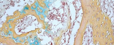
Image shows tissue derived from a single human skeletal stem cell.
Bone is in yellow, blue indicates cartilage, and marrow is pictured in red.
(CHAN AND LONGAKER et al.)
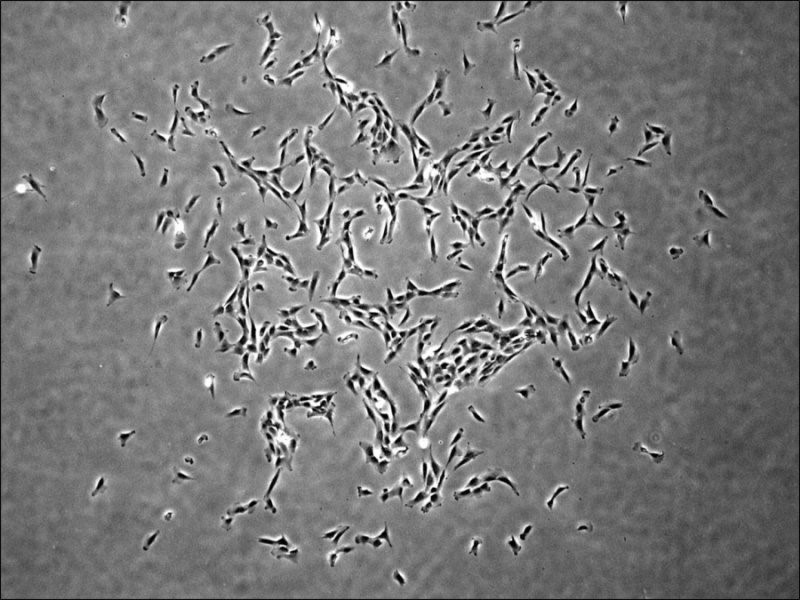
Image shows a single human skeletal stem cell-derived colony (Chan and Longaker et al.)
Human skeletal stem cells that become bone, cartilage, or stroma cells have been isolated from fetal and adult bones. This is the first time that skeletal stem cells, which had been observed in rodent models, have been identified in humans. The researchers were also able to derive the skeletal stem cells from human induced pluripotent stem cells, opening up new therapeutic possibilities for bone and cartilage related applications in the future.
Integrative detection and analysis of structural variation in cancer genomes
Jesse R. Dixon, Jie Xu, Vishnu Dileep, Ye Zhan, and Fan Song, et al. - Nature Genetics (10 September 2018)
Structural variants (SVs) can contribute to oncogenesis through a variety of mechanisms. Despite their importance, the identification of SVs in cancer genomes remains challenging. This study presents a framework that integrates optical mapping, high-throughput chromosome conformation capture (Hi-C), and whole-genome sequencing to systematically detect SVs in a variety of normal or cancer samples and cell lines.
They have identified the unique strengths of each method and demonstrate that only integrative approaches can comprehensively identify SVs in the genome. By combining Hi-C and optical mapping, and resolve complex SVs and phase multiple SV events to a single haplotype.
Furthermore, they observed widespread structural variation events affecting the functions of noncoding sequences, including the deletion of distal regulatory sequences, alteration of DNA replication timing, and the creation of novel three-dimensional chromatin structural domains. Their results indicate that noncoding SVs may be underappreciated mutational drivers in cancer genomes.
Infection of epididymal epithelial cells and leukocytes drives seminal shedding of Zika virus in a mouse model
Erin M. McDonald, Nisha K. Duggal, Jana M. Ritter, Aaron C. Brault - August 2, 2018 - https://doi.org/10.1371/journal.pntd.0006691
While Zika virus (ZIKV) is primarily a mosquito-borne virus, there are now confirmed sexual transmission cases of ZIKV from infected males to their partners. Using a previously established mouse model of sexual transmission, ZIKV was herein demonstrated to infect the testes and epididymides concurrently, suggesting that testicular infection is not required to seed infection of the epididymides. Also, replication of ZIKV was visualized by staining for ZIKV negative-strand RNA. ZIKV replication in leukocytes, immature spermatids and epididymal epithelial cells correlated with the peak of sexual transmission potential. Spermatozoa were rarely observed to stain positive for ZIKV replicative RNA intermediates, demonstrating spermatozoa are likely not a major source of infectious virus in semen. In addition, a greater fraction of the infectious virus in seminal fluids was cell-free versus cell-associated, suggesting that cell-free virus is responsible for sexual transmission. Authors provide strong evidence for epididymal epithelial cells and leukocytes, and not spermatozoa, as the major sources of infectious ZIKV in semen.
Recent advances in mass spectrometry-based approaches for proteomics and biologics: Great contribution for developing therapeutic antibodies
Noriko Iwamoto and Takashi Shimada - https://doi.org/10.1016/j.pharmthera.2017.12.007 (open access )
Mass spectrometry (MS) technologies have continued to improve dramatically, and advanced strategies that were impossible a decade ago are increasingly becoming available. The basic characteristics behind these advancements are MS resolution, quantitative accuracy, and information science for appropriate data processing.
The sequential changes of proteome expression in biological pathways are very essential, and the amounts of the changes often directly become the targets of drug discovery or indicators of clinical efficacy.
MS applications are expanding into many fields, not only basic life sciences but also forensic medicine, plant sciences, materials, and natural products. In this review, technical fundamentals and future aspects of the strategies for accurate structural identification, structure-indicated quantitation, and on the challenges for pharmacokinetics of high-molecular-weight protein biopharmaceuticals is discussed.
Gene silencing in Huntington's Disease - an exciting approach but a long way to go yet !
Leonard Wolfson Experimental Neurology Centre at the National Hospital for Neurology and Neurosurgery in London (December, 2017) Prof Sarah Tabrizi, lead researcher and director of the Huntington's Disease Centre, UCL.
Huntington's disease which is a devastating disease is caused by an error in a section of DNA called the huntingtin gene. Normally this contains the instructions for making a protein, called huntingtin, which is vital for brain development. But a genetic error corrupts this protein and turns it into a damaged toxic protein which destroys neuronal brain cells.
Studies using drug treatment designed to silence the gene is believed to work as shown below with promising results in its clinical trial - however, there is a long way to go before such a study can be medically acceptable!
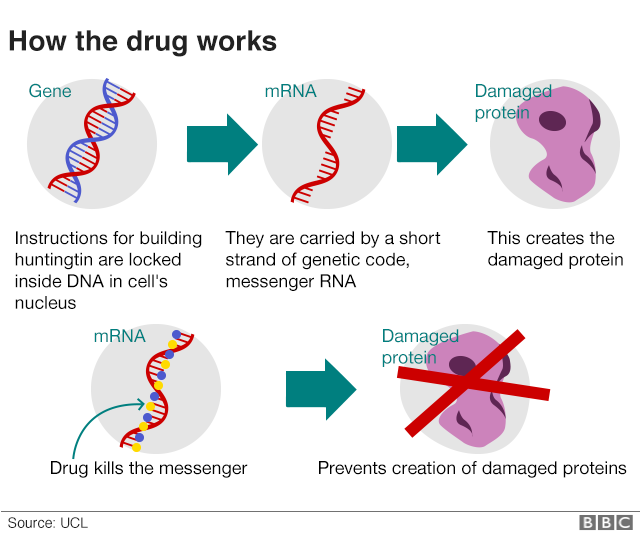
The same approach might be possible in other neurodegenerative diseases that feature the build-up of toxic proteins in the brain. The protein synuclein is implicated in Parkinson's while amyloid and tau seem to have a role in dementias. Trials are planned using gene-silencing to lower the levels of tau.
Small molecule modulation of splicing factor expression is associated with rescue from cellular senescence
Eva Latorre, Vishal C. Birar, Angela N. Sheerin, J. Charles C. Jeynes, Amy Hooper, Helen R. Dawe, David Melzer, Lynne S. Cox, Richard G. A. Faragher, Elizabeth L. Ostler and Lorna W. Harries
BMC Cell Biology: https://doi.org/10.1186/s12860-017-0147-7
This is the first demonstration that moderation of splicing factor levels is associated with reversal of cellular senescence in human primary fibroblasts. Small molecule modulators of such targets may therefore represent promising novel anti-degenerative therapies.
During the ageing process, both senescent and non-senescent cells lose a degree of response to cellular stressors. This study shows that the upstream causes of this are as yet unclear, but may include changes in genes controlling alternative splicing; a major regulator of gene expression which ensures genomic plasticity. Treatment with novel analogues of the stilbene compound resveratrol is associated not only with restoration of splicing factor expression but also with amelioration of multiple cellular senescence phenotypes in senescent human primary fibroblasts.
The precise mechanisms behind these observations are unclear, but may involve both the restoration of a more ‘youthful’ pattern of alternative splicing, and also effects of specific splicing factors on telomere maintenance. The authors suggest that the splicing factors and the upstream drivers of splicing factor expression may prove promising as druggable targets to ameliorate ageing phenotypes and hold promise as anti-degenerative compounds effective in human cells in the future.
A new approach for generating bispecific antibodies based on a common light chain format and the stable architecture of human immunoglobulin G1.
Camilla De Nardis, Linda J. A. Hendriks, Emilie Poirier, Tudor Arvinte, Piet Gros, Alexander B. H. Bakker, John de Kruif .
Journal of Biological Chemistry, 2017; jbc.M117.793497 DOI: 10.1074/jbc.M117.793497. (Sept. 2017)
A group of researchers has developed an approach to efficiently produce antibodies that can bind to two different target molecules simultaneously, a long-desired innovation in the field of cancer immunotherapy. The researchers combined different approaches - the computational tools with the biochemistry and structural biology to achieve the end results.
Antibodies are proteins produced by the immune system that specialize in recognizing and binding to molecular targets unique to bacteria, viruses or other foreign cells. Because antibodies are stable and long-lasting in the human body and can precisely recognize specific targets, they have been exploited to develop new treatments for diseases. For example, modified antibodies can be used to bind to targets in cancer cells, recruiting the immune system to attack the cancer or preventing the cancer cells from multiplying. Because of their precision and capacity to stimulate the body's immune response, antibody-based therapies typically have fewer side effects than chemotherapy or radiation.
Antibodies are "Y" shaped, and typically bind a target, or antigen, through the tip of each arm of the "Y." In naturally produced antibodies, both arms of a single antibody typically are the same and bind to the same target. One approach to increasing the versatility of antibody therapies is to engineer what are called bispecific antibodies, in which each arm binds to a different molecule. This expands the range of what antibodies can be used for. For example, a bispecific antibody could target a cluster of proteins made up of multiple protein types, or it could bring two different molecules or cell types together. Antibodies are capable of being so specific, and with bispecific antibodies, one can choose the affinities of both arms and balance them so that you can more specifically target tumors, and also recruit other cells or molecules to attack the tumor cells without many side effects.
FDA Needs Help Assessing Cutting-Edge Technologies - Nature Editorial, August, 2017
The US Food and Drug Administration (FDA) is increasingly being tasked with evaluating cutting-edge therapies and technology that its in-house expertise may not be able to address, according to a Nature editorial, which recommends academic scientists help the agency. Using the example of the 12 July 2017 meeting of the Oncologic Drugs Advisory Committee that voted unanimously in favor of the benefit-risk profile for the first of a new kind of cancer therapy, the editorial notes that much of the meeting discussed the manufacturing of the new therapy, noting that it "offers a glimpse of the regulatory complexity that cutting-edge therapies present."
FDA "needs academic scientists to get involved," the editorial adds, offering ways for them to do so via the agency’s centers of excellence and collaborations with academic institutions. "The Academics have traditionally shown little taste for the dry details of drug development. Many have fallen prey to the misconception that the FDA is not engaging with the latest science. The agency realizes that it has an image problem and, to its credit, is trying to change," the editorial says.
In addition, the editorial notes FDA’s "legendary struggle to retain its employees," particularly for "key positions such as those of bioinformaticians, where supply falls well short of demand," and as competition with industry for skilled job candidates is "particularly intense." FDA will need much more to remain effective in the face of future challenges.
Correction of a pathogenic gene mutation in human embryos: Hong Ma, Nuria Marti-Gutierrez, Sang-Wook Park, Jun Wu, Yeonmi Lee, Keiichiro Suzuki et. al.
Nature (2017) doi:10.1038/nature23305 / Published online 02 August 2017
Genome editing has potential for the targeted correction of germline mutations. Here the authors describe the correction of the heterozygous MYBPC3 mutation in human preimplantation embryos with precise CRISPR–Cas9-based targeting accuracy and high homology-directed repair efficiency by activating an endogenous, germline-specific DNA repair response. Induced double-strand breaks (DSBs) at the mutant paternal allele were predominantly repaired using the homologous wild-type maternal gene instead of a synthetic DNA template. By modulating the cell cycle stage at which the DSB was induced, they were able to avoid mosaicism in cleaving embryos and achieve a high yield of homozygous embryos carrying the wild-type MYBPC3 gene without evidence of off-target mutations. The efficiency, accuracy and safety of the approach presented suggest that it has potential to be used for the correction of heritable mutations in human embryos by complementing preimplantation genetic diagnosis. However, much remains to be considered before clinical applications, including the reproducibility of the technique with other heterozygous mutations.
CRISPR gene editing can cause hundreds of unintended mutations
Kellie A. Schafer (Stanford University), Wen-Hsuan Wu (Columbia University Medical Center), and Diana G. Colgan (Iowa) et. al - Nature Methods, May 29, 2017
As CRISPR-Cas9 starts to move into clinical trials, a new study published in Nature Methods has found that the gene-editing technology can introduce hundreds of unintended mutations into the genome. CRISPR-Cas9 editing technology—by virtue of its speed and unprecedented precision—has been a boon for scientists trying to understand the role of genes in disease. The technique has also raised hope for more powerful gene therapies that can delete or repair flawed genes, not just add new genes.
But even though CRISPR can precisely target specific stretches of DNA, it sometimes hits other parts of the genome. Most studies that search for these off-target mutations use computer algorithms to identify areas most likely to be affected and then examine those areas for deletions and insertions. These predictive algorithms seem to do a good job when CRISPR is performed in cells or tissues in a dish, but whole genome sequencing has not been employed to look for all off-target effects in living animals.
In this new study, the researchers sequenced the entire genome of mice that had undergone CRISPR gene editing in the team's previous study and looked for all mutations, including those that only altered a single nucleotide. Researchers who aren't using whole genome sequencing to find off-target effects may be missing potentially important mutations.
The power of tears: how tear proteomics research could revolutionize the clinic
Lei Zhou and Roger W. Beuerman - EXPERT REVIEW OF PROTEOMICS, VOL. 14, NO. 3, 189–191, 2017
Tear ‘omics’ research over the past decade has demonstrated the future applications of tear biomarkers for patient stratification or what is now often referred to as precision medicine. Tear collection is fast, safe, and noninvasive and offers a chance to determine the local pathology close to the disease site. The relatively simple chemical composition and sample preparation procedures make tear fluid an ideal source for diagnosis and prognosis. Proteomic studies can easily be translated into antibody-based assays for clinical use. It is hoped that a ‘tear test’ that will eventually become like a ‘blood test’ or ‘urine test’ used in eye clinics in the near future. Its application could also be extrapolated to other areas of proteomic and precision medicine.
Neuro-inflammation, neuro-protection and microglial activation
Understanding of microglial-induced neuro-inflammation is a key to understanding various neurological diseases. Microglia are the brain's resident immune cells, transitioning from resting to activation state upon sensing damage or a foreign substance. Activated microglia release a wave of chemical mediators, including chemokines, cytokines, and proteases, all of which promote the neuroinflammatory milieu. Understanding how microglia trigger 'neuro-microglial induced neuro-inflammation' may help understand the disease process, progression and the role of:
- Factors that induce microglial activation in neurodegenerative diseases
- Better understanding of ‘neuro- protection’ in the brain
Y. J. Choi et al., “Deficiency of microRNA miR-34a expands cell fate potential in pluripotent stem cells,” Science, doi:10.1126/science.aag1927, 2017.
Pluripotent stem cells are capable of generating all embryonic cell lineages but, until now, scientists could seldom manipulate induced pluripotent stem cells (iPSCs) and embryonic stem cells (ESCs) to generate extra-embryonic cell types, such as placental cells. A study published rcently (12th January,2017) in Science has now shown that removing one particular microRNA—miR-34a—from a stem cell can kick off a molecular pathway that induces endogenous retroviruses and, at the same time, enables iPSCs and ESCs to consistently form extra-embryonic cells in a dish.
The results suggest that a particular class of noncoding RNA works in concert with the latent viral elements of the genome work to limit stem cell potential, and that removing a key miRNA can lift this limitation—at least in vitro. Although stem cells can give rise to virtually any cell type inside the embryo, they have limited potential to give rise to extra-embryonic cell types. They are now trying to understand how the body restricts iPSC and ESC potential.
Exosomes
Exosomes encapsulate and transport a wide variety of molecules generated by their ‘cell-of-origin’, a process now thought to be a form of cellular signalling. Exosome signalling is common across cell types and species, but it is of particular interest in diseases with an inflammatory component. While exosome isolation and analysis is useful to understanding the mechanisms behind these multifaceted diseases, exosomes may also be exploited for their therapeutic potential. Scientists are reviewing the current knowledge on exosomes in inflammation, and exploring the potential for exosome-based therapeutics with reference to:
- The exosomal cargoes released during inflammation, and their potential as therapeutic targets
- How inflammatory diseases are uniquely suited to exosome analysis
The Cancer Genome Atlas (TCGA) project
Building on data from The Cancer Genome Atlas (TCGA) project, a multi-institutional team of scientists has completed the first large-scale "proteogenomic" study of breast cancer, linking DNA mutations to protein signaling and helping pinpoint the genes that drive cancer. Conducted by members of the National Cancer Institute's Clinical Proteomic Tumor Analysis Consortium (CPTAC), including Baylor College of Medicine, Broad Institute of MIT and Harvard, Fred Hutchinson Cancer Research Center, New York University Langone Medical Center, and Washington University School of Medicine, the study takes aim at proteins, the workhorses of the cell, and their modifications to better understand cancer. Appearing in the Advance Online Publication of Nature on May 25, 2016 the study illustrates the power of integrating genomic and proteomic data to yield a more complete picture of cancer biology than either analysis could do alone.
Antibodypedia
Systems biology, functional genomics and high-throughput proteomics efforts have massively expedited gene-product annotation and characterization. However, identifying affinity reagents suitable for secondary analyses of proteins can pose a challenge. While antibodies are widely used, their efficacy in different biological systems and experimental applications often varies depending on their immunogen or the organism in which they were produced. In fact, the likelihood that an antibody will function in a novel application or cell/ tissue type depends heavily on its affinity for the antigen and the type of epitope recognized, as well as on the antigen's concentration, folding status and post-translational modifications. Without detailed understanding of the properties of both the antibody and the biological system in which it will be used, antibody selection can require guesswork.
Antibodypedia is a searchable database of antibodies against human proteins. It aims to provide the research community with information on the effectiveness of specific antibodies in specific applications—to help scientists select the right antibody for the right application.
The Antibodypedia database was originally developed within the 6th framework EU program Proteome Binders and the project is part of the Human Antibody Initiative.
Antibodypedia contains information about publicly available antibodies generated by academic or commercial providers and directed against human protein targets; we hope to extend coverage to a range of model organisms in the near future. The database is organized in a ‘gene-centric’ manner to provide users with an overview of all antibodies available against a particular target. All antibody pages link directly to the provider for ease of access and use of the database is free-of-charge.
Precision medicine
The influence of gut microbiota on human health has been well documented, particularly in the case of metabolic disorders, such as type 1 and type 2 diabetes. In light of the strong association between the composition of one’s microbiome and human health, researchers have begun to develop targeted therapies that restore optimal balance among microbial populations. For a detailed look at the state of the microbiome and its role in precision medicine, researchers are examining:
- The human microbiome’s influence on health
- The challenges of translating microbiome studies into precision medicine therapies
A drug that heals broken bones faster and better
Author: Victoria White - Southampton University, UK
These esearchers are developing a new type of drug that may help bones heal faster and better.
Colony of human bone stem cells
Using bone samples from people undergoing hip replacement surgery, the researchers were able to show that the drug – a protein that activates a molecular pathway called the ‘Wnt’ pathway – causes stem cells found within bones to divide and to turn into more bone cells. The Wnt plays a fundamental role in animal development and disease. It is involved in controlling the growth of stem cells, which are ‘master cells’ that help restore tissues after injury. One example of this is in amphibians like salamanders. If these animals lose a leg, they can just regrow a new one.
Regenerative effect reverses if Wnt pathway switched on too long
Dr Nick Evans, Associate Professor in Bioengineering at the University of Southampton and lead author of the study, says: “Bone fractures are a big problem in society, especially in older people. It is getting worse as more people get older and their risk of fracture increases. Most fractures heal completely by themselves, but a surprising number, around 10 per cent, take over six months to heal, or never heal at all. In the worst cases this can lead to several surgical operations, or even amputation.
“Through our research, we are trying to find ways to chemically stimulate Wnt signaling using drugs. To achieve this, we selectively deliver proteins and other molecules that change Wnt signalling specifically to stem cells, particularly in the bone. This may help us find cures for many diseases, including bone disease, and speed up bone healing after fracture.”
Tumor microenvironment in tumorigenesis
The tumor milieu is the site of complex interactions between immune cells, tumor cells, and the surrounding tissue. Innovative technologies and strategies are being employed to characterize these interactions to understand the role of the tumor microenvironment in tumorigenesis and metastasis. Scientists are discussing advances in understanding of the tumor microenvironment and how this knowledge is being harnessed to develop targeted therapies with reference to:
- The role of the immune system as a mediator of the tumor microenvironment
- Modulation of the tumor microenvironment as a means of inhibiting tumor growth and suppressing metastasis
Power of microRNAs as Research Tools
microRNA (miRNA) expression provide valuable insight into disease-related transcripts, both protective and predisposing. Therapeutic design based on these data sets has enabled both supplementation with protective miRNAs such as miRNA mimics, and silencing of predisposing miRNAs using complementary RNAs. Disease-specific miRNA profiles and miRNA-based therapeutic approaches is helping researchers to understand:
- The various roles of miRNAs in disease progression and recovery
- Steps for developing miRNA-based therapeutics
|
Bioinformatics Bioinformatics is specifically premeditated with a unifying axiom providing pulpit to widen the imminent scientific creations and to enlighten Bioinformatics technologies, clinical research and computational methods. It will enable to put forth the holistic scientific approach to showcase the trends and challenges in bioinformatics. |
|
Trends and topics in Bioinformatics include: Novel technologies, Genomics & Proteomics, Computational Systems Biology, Computational Immunology, Translational & Cancer Bioinformatics, Big data & Cloud computing, Molecular & Clinical Bioinformatics, Pharmacy Informatics, Biostatistics Approaches, Neuro-Bioinformatics, Biomedical Informatics, Bioinformatics Business Trends and more. These topics are encouraged at the CEEP Conferences ! |